Metastases remain the leading cause of cancer-related mortality. At Institut Curie, CNRS and Inserm teams present new findings that follow on previous discoveries by Dr Philippe Chavrier, group leader at Institut Curie and Director of Research at CNRS 1, on the major role played by a cellular structure known as an “invadopodia” in one of the stages of the metastasis formation process, dissemination 2.
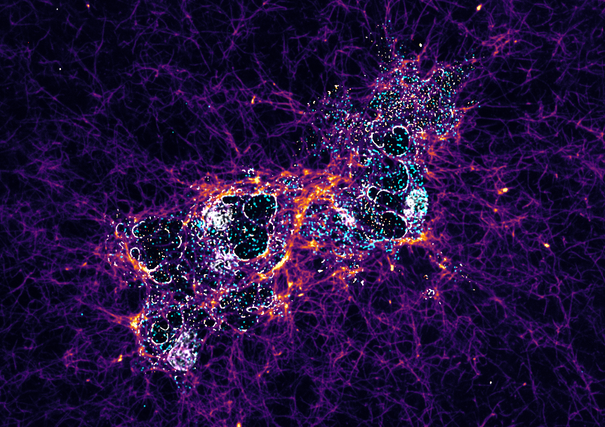
Breast cancer-derived tumor cells grown on a network of collagen fibers that mimics the tumor environment in which cancer cells are induced to disseminate during the invasive and metastatic process. The cells were labeled to reveal caveolin-1 protein, a constituent of caveolae, and TKS5 protein, essential components of invadopodia. This image reveals the close interplay between these two types of structure in breast cancer cells.
© Pedro Monteiro, Institut Curie, CNRS UMR3666 & UMR144, Inserm U1143
Caveolae and invadopodia: one structure leads to another
“In collaboration with the team of Dr Christophe Lamaze, Director of Research at Inserm and group leader at Institut Curie 3, we were originally seeking to understand the suspected role of invadopodia, the small finger-like outgrowths of tumor cells involved in the metastatic process, and their interplay with another cell structure, caveolae”, explains Dr Phillipe Chavrier, CNRS research director and group leader at Institut Curie. “Unlike the caveolae found in healthy and cancerous cells, invadopodia are only present in cancerous cells, and we believe that they were acting in tandem.”
Caveolae are recesses (invaginations) in the plasma membrane of a cell where adhesion proteins such as integrin are concentrated. They anchor the cell to its environment, namely the collagen fibers that criss-cross the extracellular matrix. The extracellular matrix is the medium in which all the cells in a tissue are immersed, enabling cell cohesion and tissue maintenance.
Pedro Monteiro, a senior post-doctoral researcher at Institut Curie, and his colleagues in the Membrane and Cytoskeleton Dynamics team have shown that when a cell becomes cancerous and form invadopodia, it does so at the point of contact between collagen fibers and the caveolae integrins where the invadopodia appear nearby, like a snap fastener. The impact of caveolae on the appearance of invadopodia is therefore referred to as “interplay”.
A coupling that promotes the dissemination of cancer cells
The caveolae are responsible for tumor invasion. The cancerous cell perceives the rigidity of its immediate environment, the extracellular matrix, thanks to the mechanical sensitivity conferred by caveolae. The invadopodia then weaken the matrix to facilitate cell mobility. The extracellular matrix, and in particular collagen fibers, is broken down by the invadopodia. As a result, the fibers become brittle and dissolve. The structural weakening of the matrix is accentuated by a mechanical force produced by the invadopodia, enabling the tumor cell to push its way through the extracellular matrix, making it possible for the cancerous cell to move. This is how the transition from the stage of tumor invasion to the spread of cancer cells takes place. Dissemination culminates in the metastatic stage, when the cells manage to spread throughout the body and find one or more new organs to invade.
Better understanding metastases: hope for therapy against cancer
This is the first study to demonstrate how the existence of this interplay between caveolae and invadopodia conditions weakening of the cell's surrounding extracellular matrix fibers. The process is described by researchers from the teams of Drs Christophe Lamaze and Philippe Chavrier as decisive in triggering the spread of cancer cells.
Although it remains to be understood how caveolae and invadopodia interact, this discovery opens up new perspectives: by targeting this mechanism, it could be possible to contain tumor cells and limit the metastatic process. This is the challenge in the search for inhibitors against the spread of cancer.
|
Reference:Pedro Monteiro, David Remy, Eline Lemerle, Fiona Routet, Anne-Sophie Macé, Chloé Guedj, Benoit Ladoux, Stéphane Vassilopoulos, Christophe Lamaze, Philippe Chavrier. A mechanosensitive caveolae-invadosome interplay drives matrix remodelling for cancer cell invasion. Nature Cell Biology (30 octobre 2023) - DOI : 10.1038/s41556-023-01272-z |
1 Membrane and Cytoskeleton Dynamics team - Cell Biology and Cancer group - UMR144 - CNRS/Université Sorbonne/Institut Curie
2 September 2020, Journal of Cell Biology (Protrudin-mediated ER-endosome contact sites promote MT1-MMP exocytosis and cell invasion), available on curie.fr: Invadopodia: How a Tumor Cell Becomes Invasive • Institut Curie
3 Membrane Mechanics and Dynamics of Intracellular Signaling - Cellular and Chemical Biology - UMR3666 / U1143 - CNRS/INSERM/Institut Curie